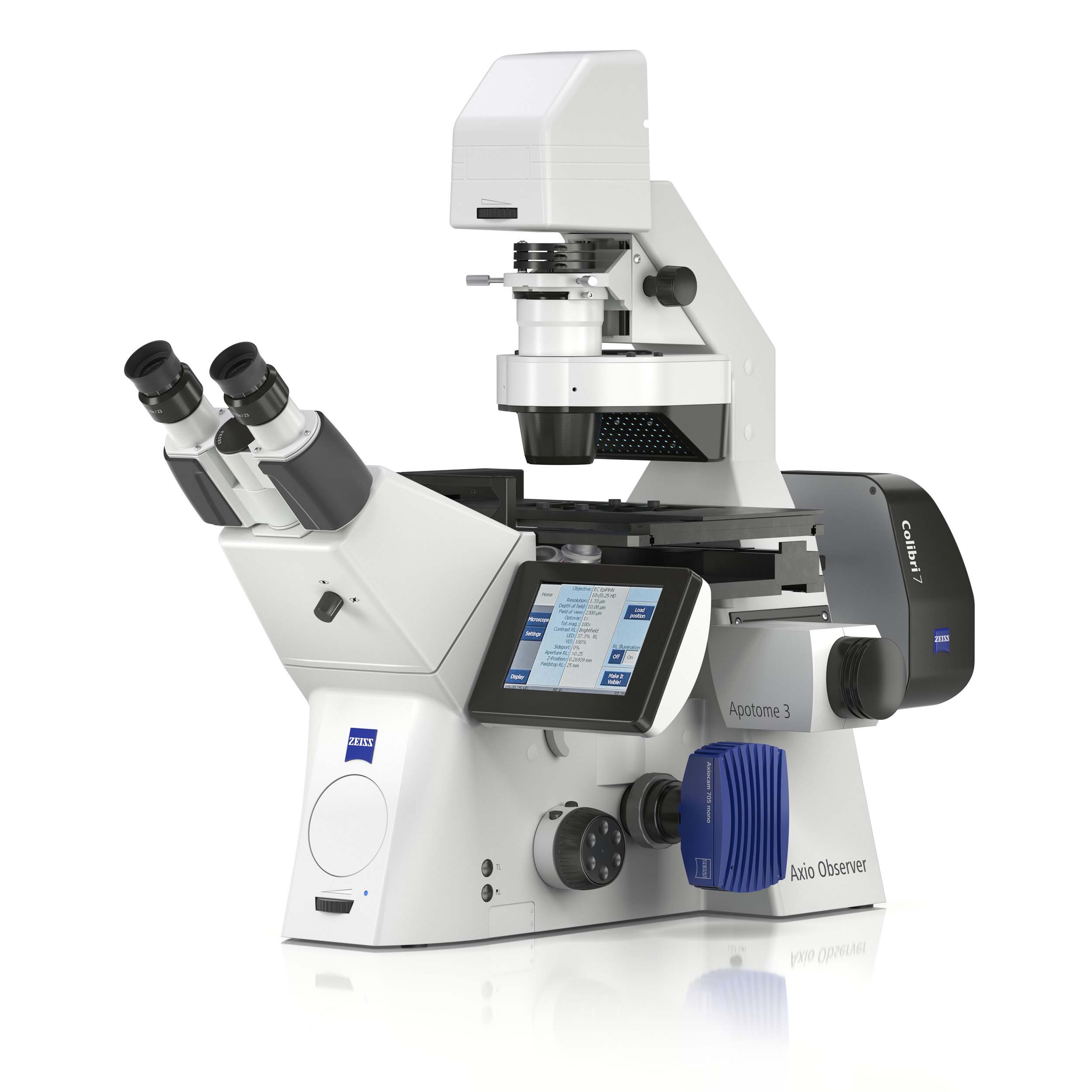
Life sciences research calls for reproducible data from a whole range of samples in a variety of conditions. ZEISS Axio Observer is your stable inverse platform for demanding multimodal imaging of living and fixed specimens, giving you:
- Ensured flexibility for technologies ranging from widefield to gentle super-resolution imaging
- Unique guidance through your experiments – from sample placement to selecting the perfect imaging modalities
- Increased efficiency thanks to a high degree of automation, economic light sources, and highest spectral flexibility
Flexibility for Your Research
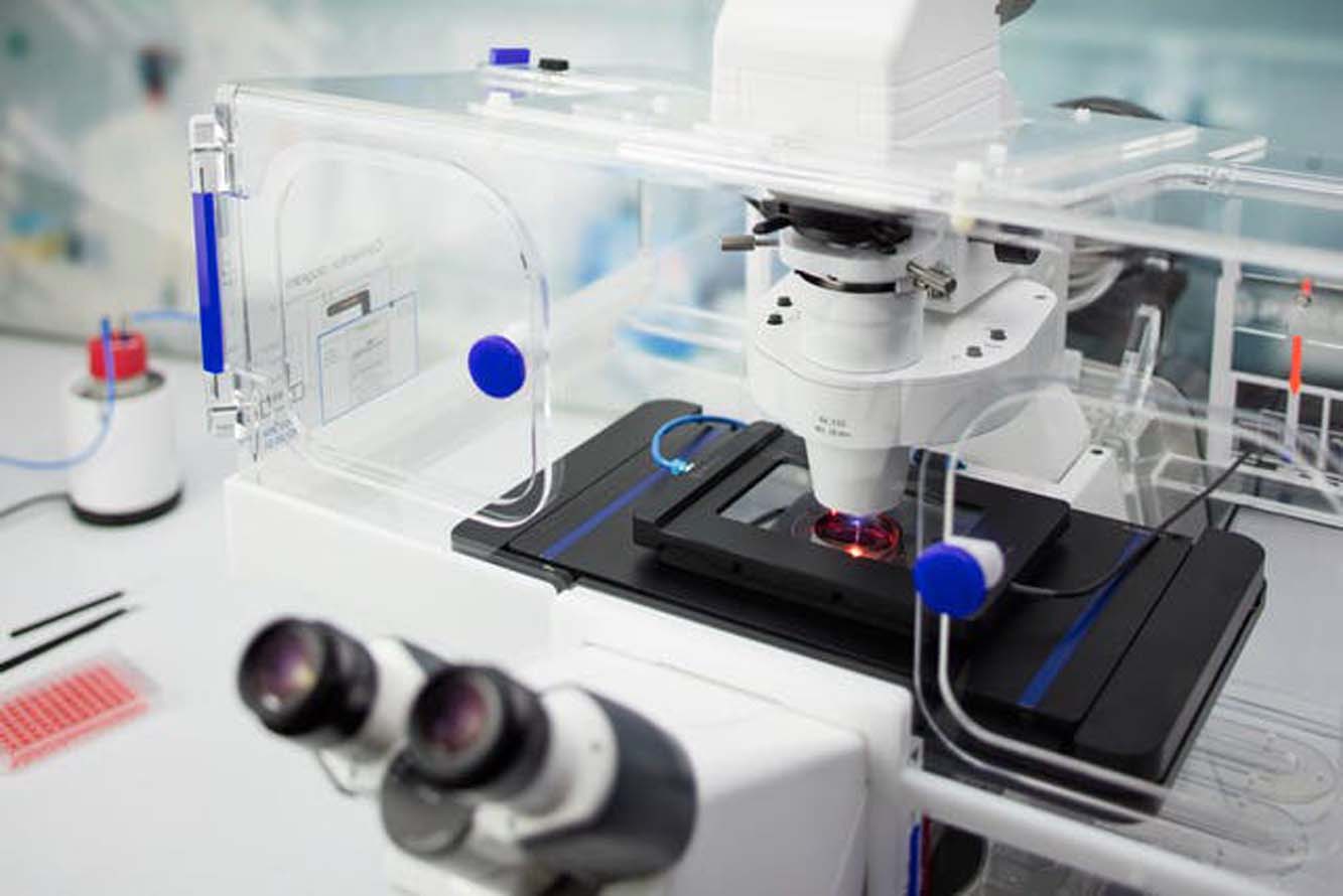
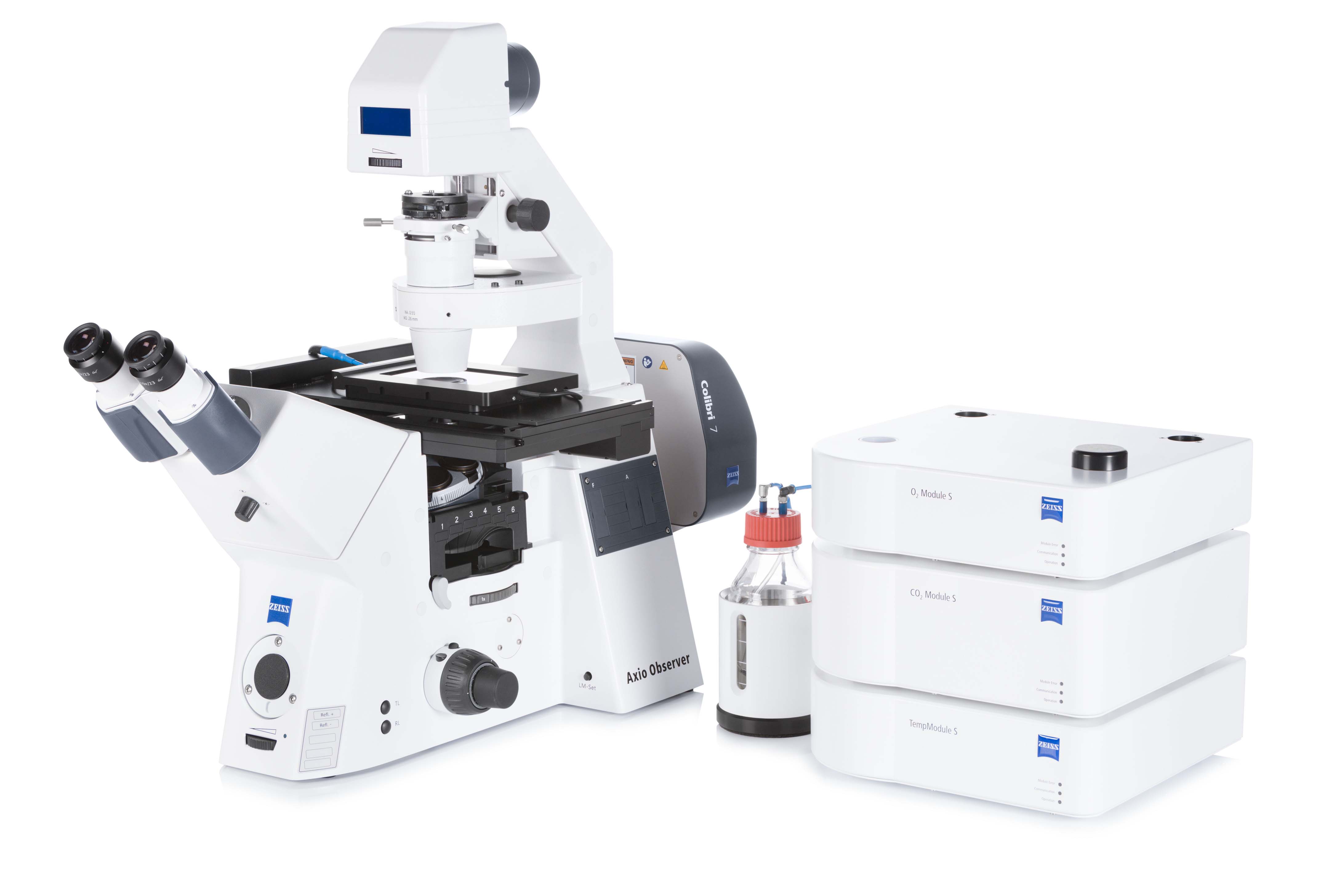
Life sciences research is a dynamic environment in which your imaging requirements are always changing. As your needs grow, Axio Observer stays with you step by step. It offers an abundance of interfaces for technologies ranging from widefield transmitted light to convenient 3D sectioning with Apotome 3, and sensitive superresolution imaging with Elyra 7 or LSM 980 and Airyscan 2. Choose the optimal incubation equipment and enjoy easy sample access for precise micromanipulation. A great variety of integrated options makes your Axio Observer both versatile now and entirely future proof.
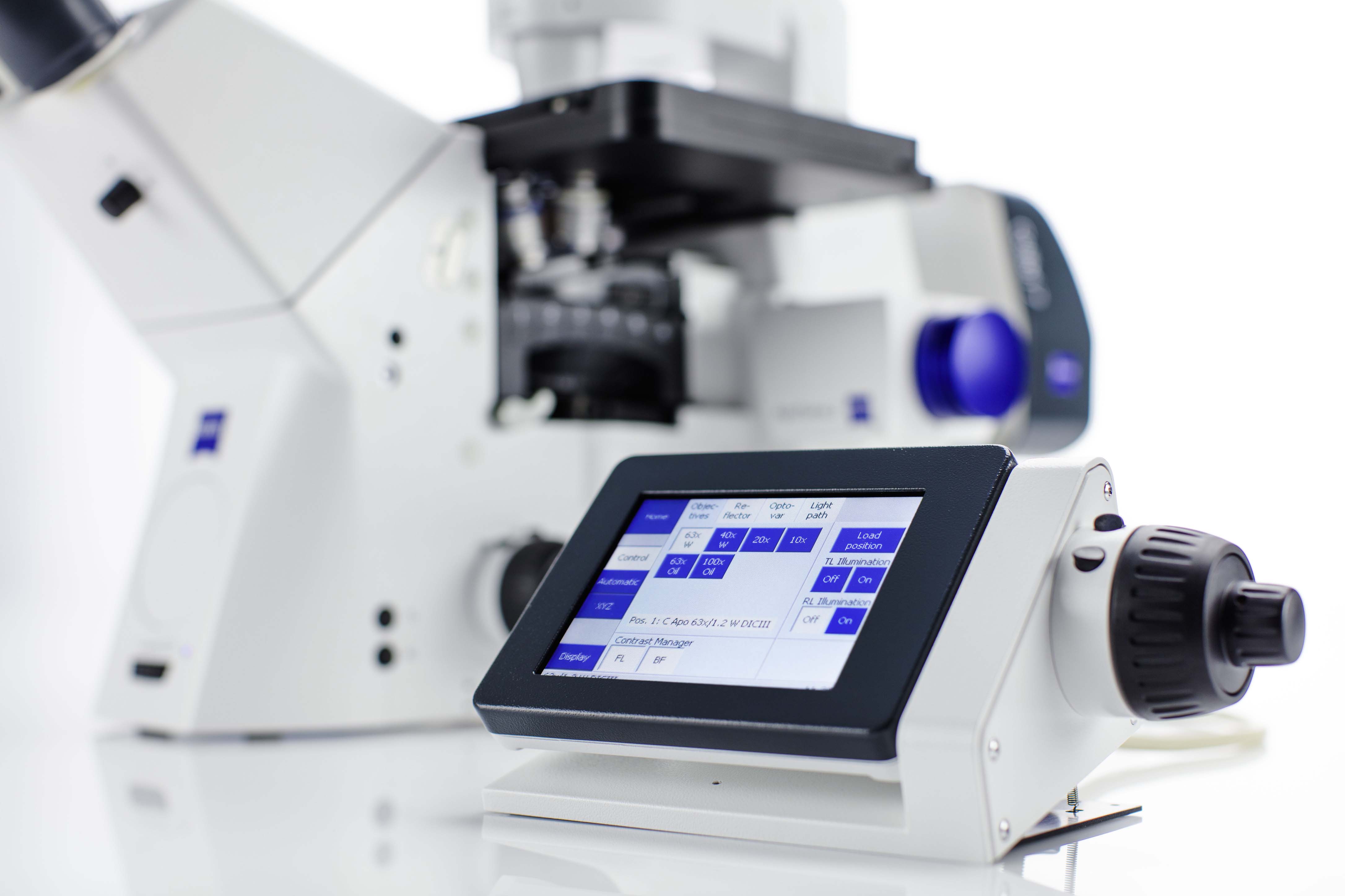
Efficiency for Your Experiments
Expect a remarkable increase in efficiency with the automation functions of Axio Observer. Use fast switchable LEDs or go for powerful and economic white-light sources in combination with the fast filter wheel for highest spectral flexibility and speed. Select the ideal camera from the dedicated ZEISS Axiocam portfolio or from third-party suppliers: You will always get the image quality and speed your applications require. With Definite Focus 3, focus drift during complicated experiments is a thing of the past. Whether keeping your sample in focus for long-term imaging or adapting your objective to your sample, it's all automatic with this highly organized system.
AI Sample Finder
Automated sample identification for efficient imaging
Microscopes are becoming increasingly automated. For sample placement, however, microscope parts such as the condenser arm often have to be moved manually. Focus adjustment and identification of the relevant areas on the sample carrier require additional manual steps. The AI Sample Finder automates this sequence, eliminating time-consuming manual adjustments and reducing the time to image from minutes to just seconds.
You can access all sample areas directly which allows you starting your experiment faster than ever. The AI Sample Finder greatly improves productivity as you can easily image only those regions containing sample not overlooking potentially important areas.
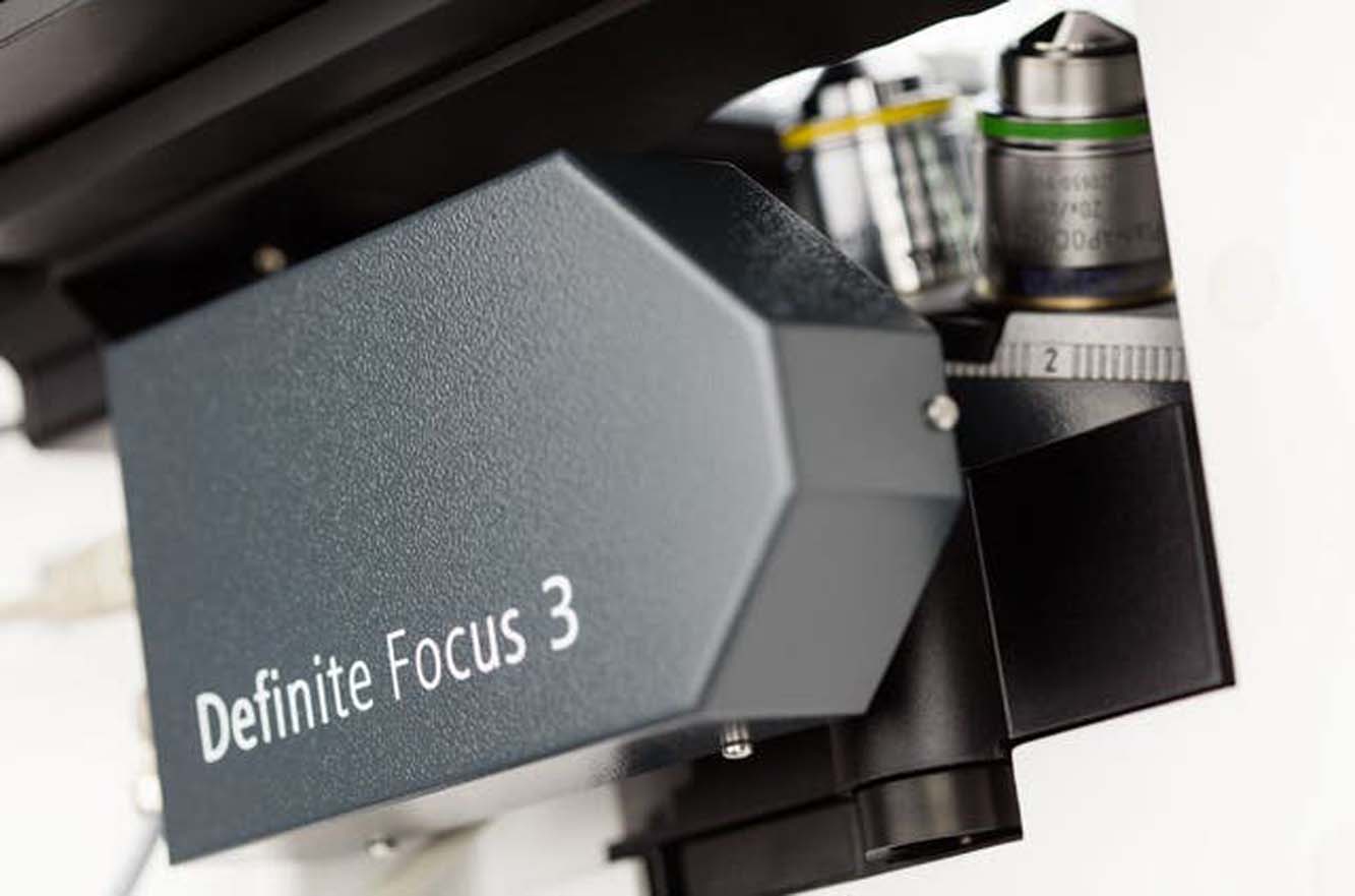
Definite Focus 3
Keep a sharp eye on your goals
Acquiring time-lapse data from living samples can be tricky. Changing conditions such as room temperature influence the microscope as well as the sample carrier and can cause focus drift. Definite Focus 3 compensates for this drift and keeps your samples in focus. With higher accuracy and precision even your most challenging multi-day, multi-position time-lapse experiments will yield sharp and high contrast images.
Here’s how it works: an infrared LED is projected through a grid onto the bottom of the sample carrier. Any change in the focal position of the sample will be indicated by a change of the grid image on the carrier bottom. An integrated camera monitors the shift while the focus drive of the stand moves to compensate for the drift in real-time. Using ZEN imaging software, simply choose a focus strategy and set up your experiment: all compensation happens automatically in the background, without interfering with your acquisition.