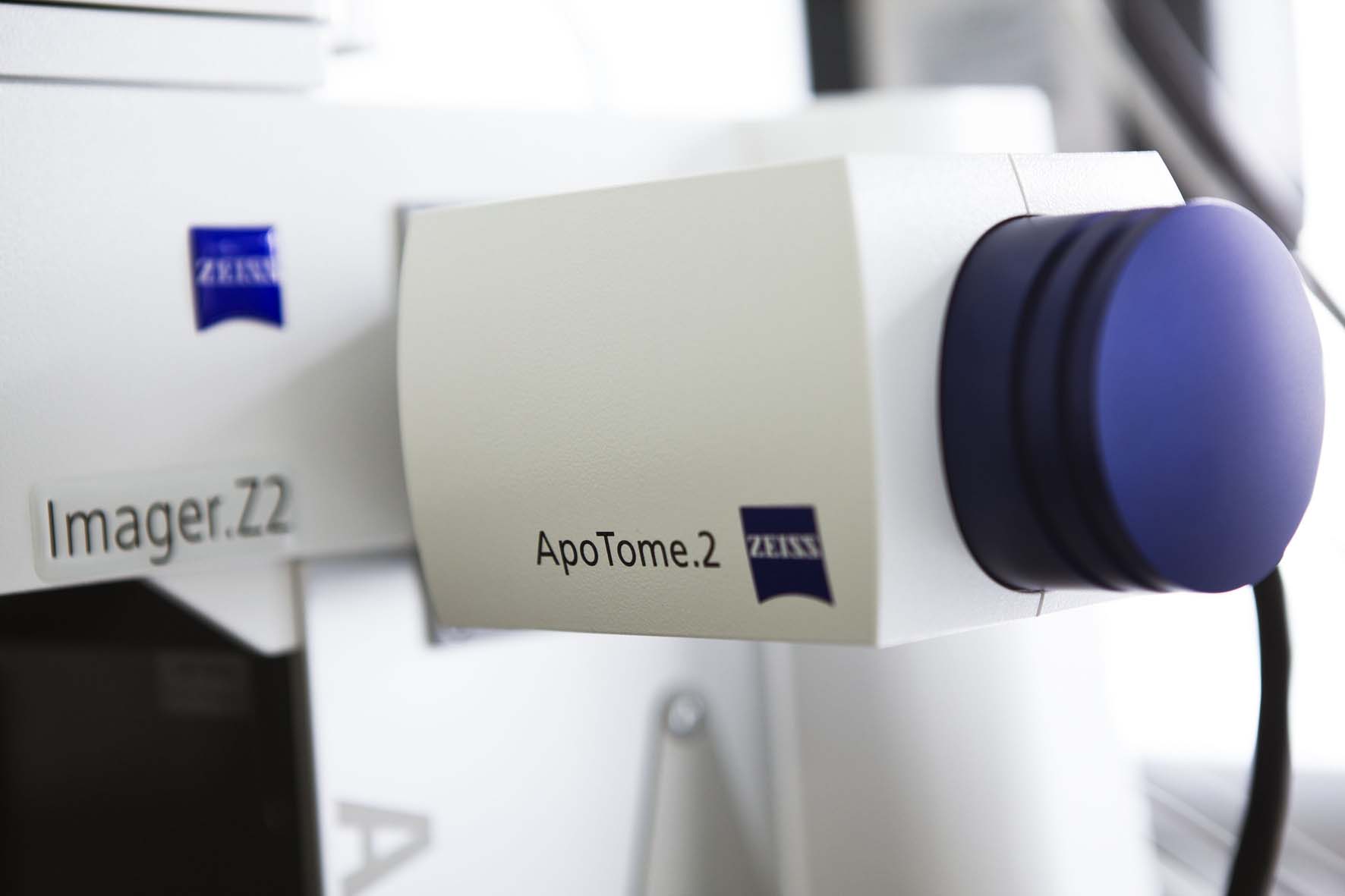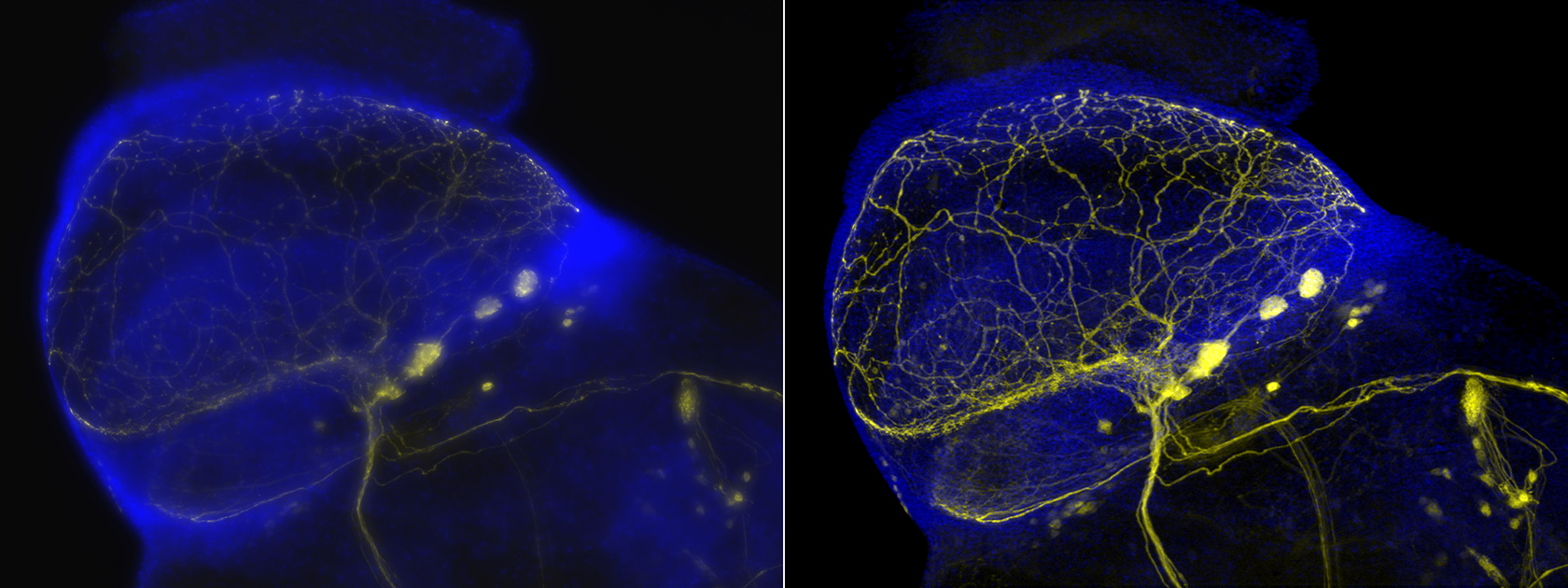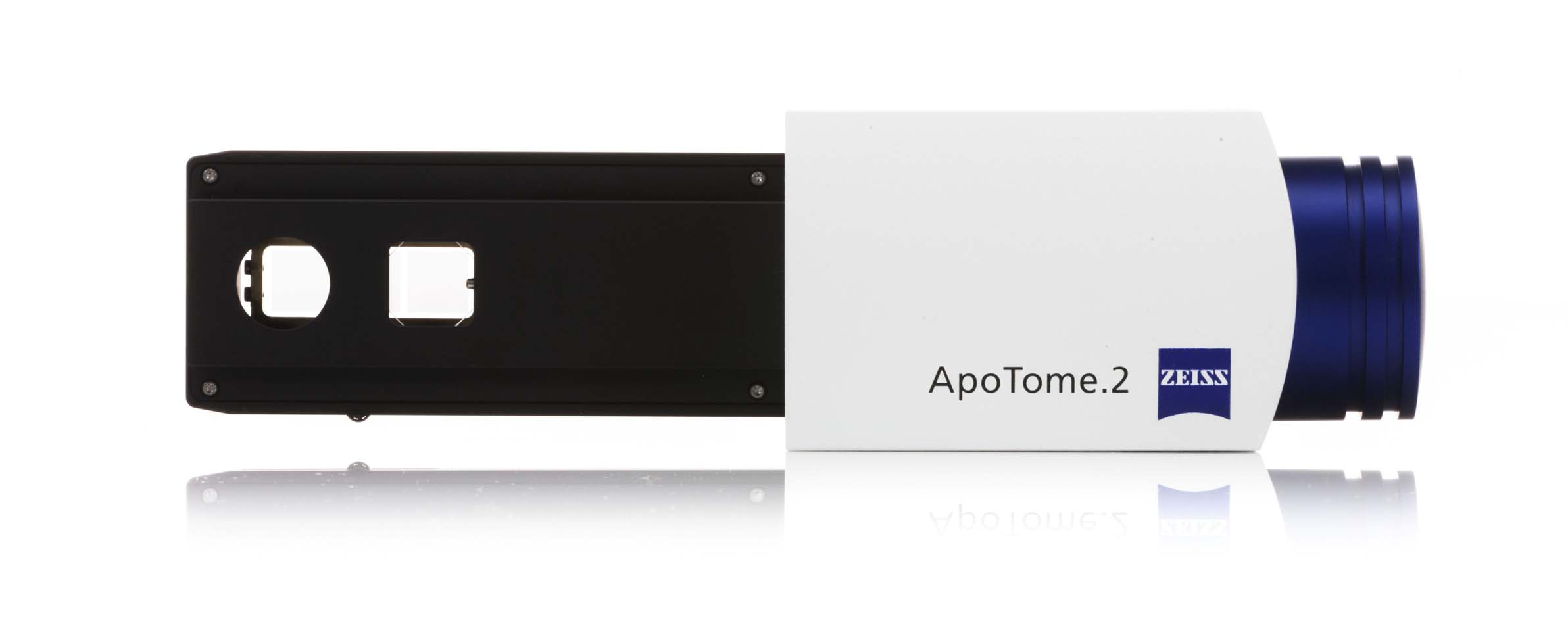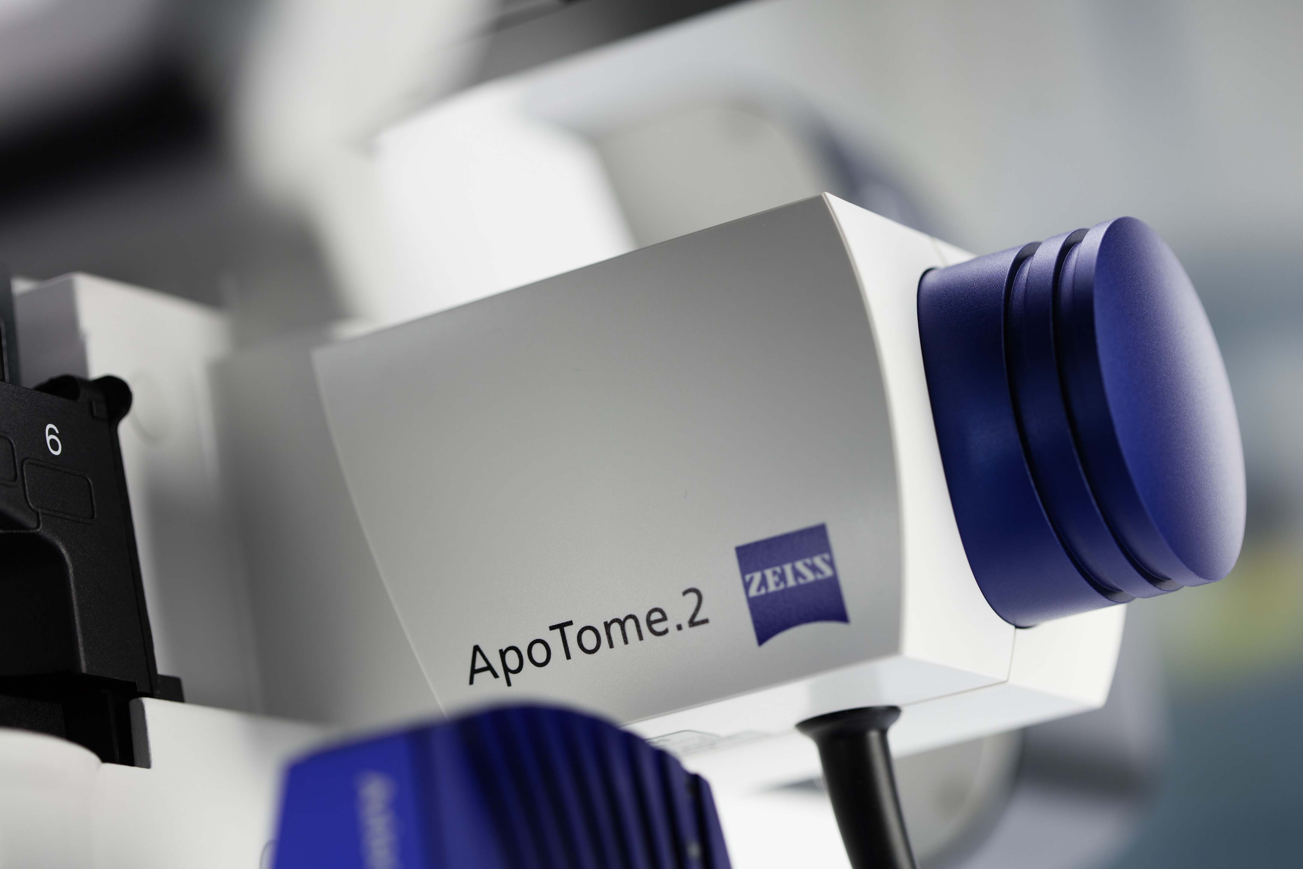


Create optical sections of your fluorescent samples - with structured Illumination, removal of out-of-focus light becomes simple and efficient, allowing you to fully focus on your research. ZEISS Apotome 3 recognizes the magnification and moves the appropriate grid into the beampath. The system then calculates your optical section from a number of images with different grid positions. It’s a totally reliable way to remove out-of-focus light, even in thicker specimens. Yet your system remains just as easy to operate as always. You get images with high contrast in the best possible resolution – simply brilliant optical sections.
To image structures of sizes ranging from hundreds of micrometers to the nanometer range, you typically use objectives with different magnifications. Apotome 3 comes with three grids of different geometries, giving you the best resolution for each objective. You can fully focus on your experiment as the ideal grid is automatically selected, always resulting in high-contrast optical sections. Apotome 3 significantly increases the axial resolution compared to conventional fluorescence microscopy: you obtain brilliant optical sections that allow 3D-rendering, even from thick specimens.
Your experiments often evolve over time in complexity and requirements. That’s why you need equipment which is not only performant but also flexible. Use Apotome 3 with conventional metal halide lamps, economic white light LEDs, or the gentle, multi-color Colibri illumination system. Simply change the filter and the system automatically moves the grid into the correct position. It’s your decision, not the technology’s: Whether you work with DAPI, Alexa488, Rhodamin, Cy5, or with vital dyes such as GFP or mCherry – Apotome 3 adapts to your fluorophores and light source, creating the sharp and brilliant images you expect.
Improve the images you created with Apotome 3 even more by deconvolution, using a patented algorithm for structured illumination. While retaining all raw data, the system allows you to switch between widefield, optical section and deconvolved images for maximum flexibility and best comparability. The fast and robust deconvolution algorithms are easy to use and improve both lateral and axial resolution of your images. Thanks to the improved contrast, higher optical resolution and suppression of existing noise, you can better recognize the structure of the examined objects.
We specialize in providing our customers with microscope and imaging solutions to meet their specific needs.
Micron Optical Ltd.
PO Box 13129, Enniscorthy,
Co. Wexford
Ireland
© Micronoptical All Rights Reserved - 2023